Cart 0

The Plasma Membrane, Through the Eyes of a High School Scientist

Author: Winnie Chan

School/Organization:

Horace Furness High School

Year: 2013

Seminar: Understanding the Process of Science and Evaluation of Evidence

Grade Level: 10

Keywords: Biology, cells, cellular transport, High School, osmosis, plasma membrane, Science, Scientific Method

School Subject(s): Biology

The plasma membrane is a thin barrier that separates the inside of the cell from its external environment. Through repeated experiments and advances in technology, scientists have developed the “Fluid Mosaic Model” to describe the structure of this dynamic membrane. Because of its unique structure, the plasma membrane can effectively regulate what enters and exits the cell. Students need to understand how the plasma membrane works in order to fully grasp the concepts the come later on.

This unit is designed to teach 9th and 10th grade biology students about the content using general principles of scientific processes learned in the beginning of the year. Students will explore the plasma membrane and cellular transport through the eyes of a scientist learning about this topic for the first time. Students will plan and carry out experiments, analyze and interpret data, and evaluate and communicate the information learned.

Download Unit: 13.02.03-unit.pdf

Did you try this unit in your classroom? Give us your feedback here.


Full Unit Text
Rationale

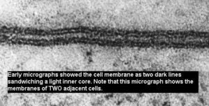
The fluid mosaic model of the plasma membrane developed as a result of evidence gathered from conceptual and technological advances. In the late 1800s, scientists initially hypothesized that membranes must be made of lipids, after observing that substances dissolved in lipids entered cells more quickly than those insoluble in lipids (Campbell 138). After experiments with red blood cells, two Dutch scientists, E. Gorter and F. Grendel, proposed that membranes must actually be a phospholipid bilayer, two molecules thick. This bilayer would provide a stable boundary between the aqueous environments inside and outside the cell.

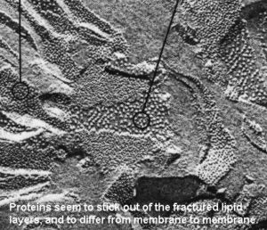
With the development of the electron microscope, freeze-fracture methods and additional studies with capillaries, scientist were able to further conclude that proteins were found embedded within the plasma membrane (integral proteins) or along the sides (peripheral protein).

Continued research eventually led to the most current model of the plasma membrane, one that is composed of a phospholipid bilayer embedded with cholesterol molecules, cytoskeleton, and long carbohydrate chains, in addition to protein molecules. The phospholipid bilayer contains both hydrophobic and hydrophilic regions, as do many of the integral proteins themselves.

The plasma membrane plays a role in structural support, cell to cell communication and cell identification. However, its’ main function is to controls what enters and exits the cell. Because of its unique structure, only certain substances are allowed to freely enter and exit the cell. For this reason, the plasma membrane is “semi-permeable”.

Passive cellular transport is diffusion, the tendency for molecules to spread out into the available space, across a membrane (Campbell 145). Molecules will move down its concentration gradient, to areas of lower concentration, in order to reach equilibrium. Some small, non-polar molecules (such as carbon dioxide and oxygen gas) can simply pass through the plasma membrane. Other small, polar substances (such as sodium and potassium ions) must pass through a protein channel in order to enter or exit the cell (Holt 80).

The passive transport of water across the plasma membrane is called osmosis. In osmosis, water moves towards areas of higher solute concentration because the solute particles are too large in size to move across the membrane. Cells can be found in one of three solutions in varying concentrations. Hypertonic solutions contain more solute than the cell itself. If a red blood cell is placed in a hypertonic solution, for example, water will move out of the cell and into the solution, causing the cell to decrease in size. Hypotonic solutions contain a lower solute concentration than the cell. Isotonic solutions are those equal solute concentration as the cell (Campbell 146).

Students often have difficulty understanding this portion of the biology curriculum because the vocabulary is new, the content is dense and they often struggle to relate the information to their own background knowledge. Explicit teaching of the plasma membrane and cellular transport leads to rote memorization of facts, which tend to be quickly forgotten moments after a unit assessment. Student learning must be an active process that connects to problems relevant to students’ lives (Chaplin and Manske, 2005). By challenging students to make connections beyond just words on a page, they will have better memory recall of how the plasma membrane works, which will then help them understand the concepts that come later (i.e. how glucose enters the cell in cellular respiration, how viruses are able to recognize and attach to particular cells, how eggs and sperm fuse during fertilization).

Objectives

The objectives of the unit will include the following:

  • Correctly draw and label the components of the plasma membrane
  • Analyze the relationship between the structure and function of the plasma membrane
  • Determine what factors will impact whether a substance will pass through a membrane
  • Explain mechanisms organisms use to maintain homeostasis
  • Compare different types of molecular transport
  • Interpret data and draw conclusions
  • Obtain, evaluate and communicate information in writing

Strategies

This unit will include a variety of lessons that will cater to different learning modalities. The unit will start with inquiry based lessons and activities that will guide students to draw their own conclusions about plasma membrane structure and function.


Instead of a teacher-centered lecture about the plasma membrane composition, students will be given data from early experiments and asked to think about what this information tells us about the membrane. Based on the information learned, students will build a 3D model of the plasma membrane in structured groups of 3-4.


After students understand the membrane structure and composition, they will conduct a series of experiments exploring osmosis and cellular transport. Through a series of guided questions and observations, students will discover on their own how substances are moved across the membrane. Teachers reinforce the major points of cellular transport after the experiments are complete. Finally, students will apply their knowledge by working in small groups to complete a case study on osmosis.

Classroom Activities


The proposed unit is for a ninth grade biology course. The class meets every day for 51 minutes. At Horace Furness High School, science classes have access to a lab room and full-time lab assistant one day a week.


Students should have sufficient background knowledge on water properties (including polarity, hydrophobic/hydrophillic terminology) and macromolecules (especially phospholipids and proteins) going into this unit.


Lesson One

– Objective: Correctly state the components of the plasma membrane and describe/show how they are organized in the plasma membrane

– Activities:

  • Students will be given a 2-sided handout that they are to complete in order. The front page summarizes data from an early plasma membrane experiment (see Appendix for Handout 1: “Lipid Layer Evidence”). Students will calculate the ratio between total surface area and lipid surface area in the red blood cells of various animals. The resulting 2:1 ratio shows that the plasma membrane must be 2 lipid molecules thick.
  • The last question on the front page will ask students to sketch how they think the plasma membrane looks, knowing that (i) the membrane is composed of proteins and 2 lipid molecules (ii) the space inside (cytoplasm) and outside the cell are polar.
  • The back page will present students with the freeze fracture electronmicrograph images. They will propose possible explanations to a series of guided questions leading them to deduce that proteins are embedded throughout the plasma membrane.
  • The lesson will wrap up with 2 exit slip questions: (1) List the macromolecules present in the cell membrane. (2) How do we know that the plasma membrane must include a lipid bilayer?


Lesson Two

– Objective: Construct the most current fluid mosaic model of the plasma membrane using a variety of supplies.

– Activities:

  • Students will watch a TEDEducation video reviewing the Gorder and Grendel experiment: “Insights into Cell Membranes via Dish Detergent” (http://ed.ted.com/lessons/insights-into-cell-membranes-via-dish-detergent-ethan-perlstein).
  • The teacher will screen (or distribute paper copies per table of) a timeline of the various proposed models of the cell membrane and explain how the model changed over time as technology advanced. For a copy of the timeline, go to Sheet B 1.2 at: http://www.nuffieldfoundation.org/sites/default/files/files/resources%20part%201_cell%20membranes.pdf
  • Students will work in small assigned groups to construct the most current fluid mosaic 3D model of the plasma membrane. Each group of 3-4 students will be given the following: one handout that shows the fluid mosaic model in 2D form (see Handout Two: Fluid Mosaic 3D Model), 50 cotton swabs, rubber band (to hold the cotton swabs together), pipe cleaners, straws cut in half, tiny twizzler candy pieces, one marble and one grain of rice. Students are to build the model given the supplies and then answer the questions on the handout.
  • The lesson will wrap up with the teacher drawing out a (simpler) 2D version of the Fluid Mosaic model that students copy (and subsequently label) into their notebooks. The teacher guides a concluding discussion on why the plasma membrane is called a Fluid Mosaic Model.
  • Homework: Students are given a copy of the lab handout “Diffusion Through a Selectively Permeable Membrane” (http://serendip.brynmawr.edu/sci_edu/waldron/#diffusion)Students are instructed to:
    • Answer 4 of the 5 “starred” questions on pages 1 and 2.
    • Read the procedure to the lab.


Lesson Three:

Objective: (1) Analyze the relationship between structure and function of the plasma membrane (2) Determine what factors will impact whether a substance will pass through a membrane

Activities:

  • The lesson begins with a review of the plasma membrane model built the day before. The teacher projects a hand-drawn picture of the model and three questions: (i) Why can’t the rice easily pass through the Q tip? (ii) Why can CO2 gas easily pass through? (iii) Why can’t the marble easily go through? (iv) What is the primary function of the plasma membrane? As a class, the student and teacher label the picture and answer the questions; different students will come up to the board to record the answers.
  • Students complete pages 2, 3 and 5 in the lab “Diffusion Through a Selectively Permeable Membrane”.
  • Homework: Students finish up their lab handout.
  • Teacher note: Prepare for the “Modeling Passive Transport with an Egg Cell Lab” before leaving school for the day. Soak eggs in plastic cups filled with vinegar for at least 24 hours. You will need enough eggs so that each group of 3-4 get two eggs.


Lesson Four:

Objective: Determine what factors will impact whether a substance will pass through a membrane

Activities:

  • The lesson begins with a review of yesterday’s lab (the teacher shows the set-up and results to the class). Students then form groups of 3-4 and go over their answers to the “Conclusion” questions.
  • With the class, the teacher diagrams the 2 types of passive transport illustrated in class so far. Students copy the diagrams into their notebooks. Below their pictures, students write 2 requirements needed for each type of transport.
  • In the final 25 minutes of class, students will begin the “Modeling Passive Transport with an Egg Cell Lab”.
    • Begin the lab by explaining: (i) why vinegar dissolves the shell of an egg (ii) what the egg represents
    • Students will pour out the vinegar in each cup, find the mass of each shell-less egg, record the information onto their lab handout and fill one cup with corn syrup, the other with vinegar. See the appendix for Student Handout Three: “Modeling Passive Transport with an Egg Cell Lab Guidelines”.
  • If there is extra time after the egg lab set-up, the teacher can review with the students the purpose and format of a science lab report.


Lesson Five:

Objective: Explain mechanisms organisms use to maintain homeostasis

Activities:

  • Students rinse out the eggs in corn syrup and vinegar. They make their observations, find the mass of both eggs and record them in the data section on their lab handout. Students work in their lab groups to answer a series of guided questions (on the lab handout) that will help them understand why water has moved into and out of the egg cell, not sugar or acetic acid.
  • In the final 30 minutes of class, the teacher will review the purpose and format of a science lab report, as well as what an introduction of a science lab report should contain.
  • Students begin working on the introduction to their lab report.
  • Homework: Students finish the introduction to their lab report.


Lesson Six:

Objective: Write informative/explanatory texts to examine and convey complex ideas, concepts, and information clearly and accurately

Activities:

  • The lesson begins with the teacher reviewing the significance of a lab report and what belongs in each section.
  • Students will work on their lab report for the duration of the period. What they do not finish in class will be completed for homework. Lab reports are typically due three days after the day of the lab.


Lesson Seven:

Objective: Explain mechanisms organisms use to maintain homeostasis

Activities:

  • Students will read the Part II of a case study on osmosis (“Osmosis is Serious Business”) and answer the corresponding questions. A copy of this case study can be found at: http://sciencecases.lib.buffalo.edu/cs/files/osmosis.pdf
  • In the final fifteen minutes of class, students will partner up and discuss their answers to the questions. In pairs, the students must also come up with a picture that illustrates what happened to the patients’ cells as a result of the IV drip. The teacher will circulate the room and listen to the discussions, adding additional questions/commentary as needed.
  • Homework: Read Part I of the case study and answer the corresponding questions.


The unit will culminate in a written exam in which students demonstrate their knowledge by answering multiple choice and free response questions. Many of these questions will be pulled directly from a sample high-stakes exam (i.e. Biology Keystone, Biology SAT, ACT Science and/or Science PSSA).

Annotated Bibliography

Student Resources


Doherty, Jennifer, and Ingrid Waldron. “Diffusion Across a Selectively Permeable

Membrane.” Hands-on Activities for Teaching Biology to High School or

Middle School Students. N.p., n.d. Web. 14 June 2013.


[This is a very clear and straightforward passive transport lab handout for students.]


Johnson, George B. Holt Biology. Orlando, Fl.: Holt, Rinehart and Winston, 2004. Print.


[This biology textbook provides a good reference and pictures for students.]


“Lesson B: Cell Membranes.” Nuffield Foundation Teaching About Science. N.p., n.d.

Web. 05 Mar. 2013.


[This website provides experimental data on the discovery of the Fluid Mosiac Model of the plasma membrane.]


Nash, Troy. “Osmosis Is Serious Business.” National Center for Case Study Teaching

in Science (NCCSTS). N.p., n.d. Web.

<http://sciencecases.lib.buffalo.edu/cs/files/osmosis.pdf>.


[An interesting case study relating osmosis to plant and human health.]


Teacher Resources


Campbell, Neil A., and Jane B. Reece. Biology. San Francisco: Benjamin Cummings,

  1. Print.


[This college biology textbook is a great resource on all science topics.]


Chaplin, Susan B. “A Theme-Based Approach to Teaching Non-Majors Biology.”

NSTA.org. N.p., Sept. 2005. Web.


[This journal article describes another approach to making science learning more relevant to students.]


Models of the Plasma Membrane. N.p., n.d. Web.

<http://www.hsc.on.ca/moffatt/bio3a/cellbio/phase1.htm>.


[This website outlines how the models of the plasma membrane have changed over time.]


Perlstein, Ethan. “Insights into Cell Membranes via Dish Detergent.” TEDEducation,

n.d. Web. 30 Mar. 2013.


[This video is a great introduction to the composition of cell membranes.]


Pike, Angela. “Build A Model of the Cell Membrane.” Education.com. N.p., n.d. Web.

<http://www.education.com/science-fair/article/build-cell-membrane-model/>.


[This building activity is a great way for students to visualize the cell membrane.]


Classroom Materials


Additional materials required for this unit include:

– Cell membrane model supplies per group of 3-4 students: 50 cotton swabs, rubber band (to hold the cotton swabs together), pipe cleaners, straws cut in half, tiny twizzler candy pieces, one marble and one grain of rice

– A list of supplies needed for “Diffusion Across A Selectively Permeable Membrane” can be found at: http://serendip.brynmawr.edu/sci_edu/waldron/pdf/MembraneTeachPrep.pdf

– Egg Cell Lab supplies per group of 3-4 students: 2 eggs, 2 plastic cups, 1 toothpick, masking tape (to label the plastic cups), vinegar and corn syrup.

Appendix

Handout One: “Lipid Layer Evidence”


Front page:


Background:

Data from the experiment which laid the foundations for a model of membrane structure is summarized in the table below. Gorter and Grendel obtained the

membranes of red blood cells. They calculated the area of the red blood cell

membrane and then extracted the lipids that were present. These lipids were dissolved in petroleum ether and allowed to spread into a layer one molecule thick on a surface of water and the area was measured.


**Remember that “surface area” is the total area of a 3D object.


Directions:

Divide ‘B’ by ‘A’ to determine the ratio of total surface area to lipid surface area. Then, answer the analysis questions that follow.


Animal Total surface are of the red blood cell (A) Surface area occupied by the lipids extracted (B) Ratio

B/A

Dog 31.3 62
6.2 12.2
Sheep 2.95 6.2
2.65 5.8
Rabbit 5.46 9.9
5.46 8.8
Guinea-pig 0.52 1.02
0.52 0.97
Goat 0.33 0.69
0.33 0.63
Man 0.47 0.92
0.47 0.989


Analysis Questions:

  1. Why do you think Gorter and Grendel used more than one red blood cell per organism?
  2. Lightly shade in the surface area of the red blood cell cut in half.

  1. The ratio of total surface area to lipid surface area is ________.
  2. Circle the correct number: “The red blood cell must be covered by a lipid layer that is

[ 1   2   3 ] molecules thick.

  1. Follow-up experiments also determined that proteins were present in the plasma membrane, in addition to lipids. In the space below, sketch how you think the molecular view of the plasma membrane might appear. Label your sketch! Keep in mind that the space inside the cell (cytoplasm) and outside the cell are polar.


Back page:

  1. The micrograph above shows the membranes of TWO adjacent cells. What do you notice about the membranes?

  1. Using “freeze-fracture” techniques, biologists were able to split cell membranes along the lipid layer. In the image above, you can see the inside of some membranes (those that have been circled). Describe what you see inside the membrane.


  1. These bumps sticking out were variable from membrane to membrane, and were about the right size to be proteins. From these pictures, do you think scientists concluded that proteins were found sandwiched on top of the lipid bilayer or embedded within?


  1. Using the knowledge you’ve learned so far, draw how you think the plasma membrane looks. Keep in mind that the inside and outside environments of the cell are aqueous.


Handout Two: Fluid Mosaic 3D Model


Follow the following procedure to make a model of the plasma membrane. When you come across a question, answer it in complete sentences.

  1. Gather the cotton swabs into a bundle and place the rubber band around the middle to keep them in a bundle.
  2. Place a receptor molecule into the cell membrane.
    1. Take one of the pipe cleaners and place it through the bundle of cotton swabs.
    2. Bend one end of it into a circular shape. This shape represents how signal molecules bind to specific molecules. Only a circular-shaped molecule can bind with this receptor.
  3. Use the second pipe cleaner as a carbohydrate chain. Place it in the bundle of cotton swabs, just as in step 2. Don’t bend this pipe cleaner.
  4. Cut your drinking straw in half. Place each half into different locations in the bundle of cotton swabs. These represent the protein channels and pumps.
  5. Holding the cotton swabs vertically, place the marble on top of the swabs. Does it pass between the swabs? Why or why not?
  6. Place the marble on top of the straw. Does it pass through the straw? Why or why not?
  7. Still holding the cotton swabs vertically, place the grain of rice on top of the swabs. Does it pass between the swabs? Why or why not?
  8. Place the grain of rice on top of the straw. Does it pass through the straw? Why or why not?
  9. Place your mouth on the cotton end of the swabs and blow. Can you feel air (carbon dioxide gas) on the other side of the swabs? Why or why not?
  10. Explain how the swabs and straws actually represent the components of the a real cell membrane.
  11. Roll the bundle of cotton swabs between your hands. Do the individual swabs move? Without pulling the straw out can you move it between the swabs? How does this represent the fluid mosaic model?

Handout Three: “Modeling Passive Transport with an Egg Cell Lab Guidelines”


Introduction: The purpose of this lab is to soak a shell-less egg in various liquids overnight and observe how the size of the egg changes. The shell-less egg will represent a cell and its selectively permeable membrane. Eggshells are made up of the mineral calcium carbonate, which dissolves in acids such as vinegar. Your eggs have been soaked in vinegar overnight. After the shell has been dissolved, only the membrane will remain around the egg.


Pre-Lab Questions:

  1. Draw the current model of the plasma membrane in the space below.


  1. Cell membranes are selectively permeable. What does this mean?


  1. Read the introduction paragraph above. What is the purpose of this lab?


  1. How was the eggshell removed?


  1. What does the shell-less egg in this lab represent?


  1. Make a hypothesis! Predict what will happen to the size of the egg when it is soaked overnight in:
  2. Corn syrup
  3. Vinegar (5% acetic acid, 95% distilled water)


Table 1:

Liquid Beginning mass (g) Mass after soaking (g) Observations (written and pictorial) Percent Mass Change
Corn syrup


Vinegar (5% acetic acid, 95% distilled water)


Analysis Questions:

  1. Were your predictions correct? Which liquid caused the egg to swell? Which caused the egg to shrink?


  1. When you poked the vinegar egg with a toothpick, what came out? When you poked the corn syrup egg with a toothpick, what came out?


  1. Why do you think the egg in vinegar swelled in size? Why do you think the egg in corn syrup shrank? Read your answer to analysis question 2 before you answer this question!

Content Standards

This unit aligns to the Pennsylvania Academic Standards for Science and Technology and Engineering Education.


3.1.A

  • Explain how the cell membrane functions as a regulatory structure and protective barrier for the cell.
  • Describe transport mechanisms across the plasma membrane.
  • Formulate and revise explanations and models using logic and evidence.
  • Recognize and analyze alternative explanations and models.
  • Evaluate experimental information for relevance and adherence to science processes.
  • Judge that conclusions are consistent and logical with experimental conditions.
  • Interpret results of experimental research to predict new information, propose additional investigable questions, or advance a solution.